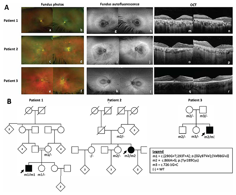
Figure 1. Clinical findings and family pedigrees for patients 1-3.
(A) Optos pseudocolour fundus photographs (a-f), Optos fundus autofluorescence (g-l) and Spectralis optical coherence tomography (OCT) images (m-r) for patients 1-3 with biallelic SUMF1 variants and retinal dystrophy (B) Family pedigrees showing genotypes and familial segregation where available.

