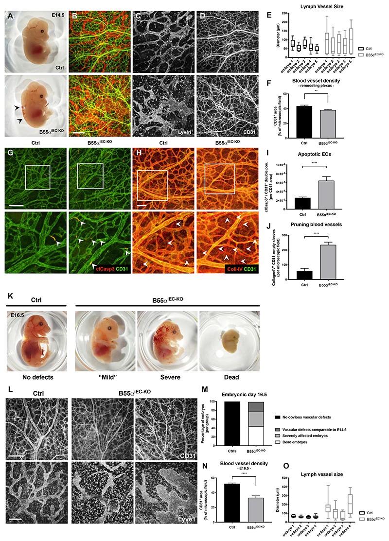
Figure 2. Endothelial loss of B55α leads to severe vascular and lymphatic defects during embryonic development resulting in embryonic lethality.
(A) Representative images of E14.5 embryos lacking endothelial B55α and littermate controls. (B-D) Whole mount skin samples stained for lymph vessel marker Lyve1 (C) and the blood vessel marker CD31 (Scale bar: 200µm) (D)
(E,F) Quantitative analysis of lymph vessel diameter (n = 4 embryos per group) (E) and blood vessel density in the remodeling vascular plexus (n ≥ 12 embryos per group) (F).
(G,I) Apoptotic ECs in the remodeling vascular plexus indicated by immunostainings against CD31 and cleaved Caspase3 (Scale bar: 100µm) (G) and quantitative analysis thereof (I).
(H,J) Pruning blood vessels, visualized by Co-staining of CD31+ and CD31- / CollagenIV+ empty sleeves (Scale bar: 100µm) (H) and quantitative analysis thereof (n = 4 per group) (J).
(K,M) Representative images of whole embryos at stage E16.5 showing differentially affected mutants and statistical quantifications thereof (M).
(L,N,O) Representative images of embryonic skin samples stained for CD31+ blood vessels and Lyve1+ lymph vessels (L) and statistical analysis thereof (Scale bars = 200µm) (N,O) The statistical analysis was performed on 9 embryos per group for (N) and 4 embryos per group for (O).
P values are *p<0,05; **p<0,001; ***p<0,001; ****p<0,0001.
Graphs show standard error of the mean (SEM).

