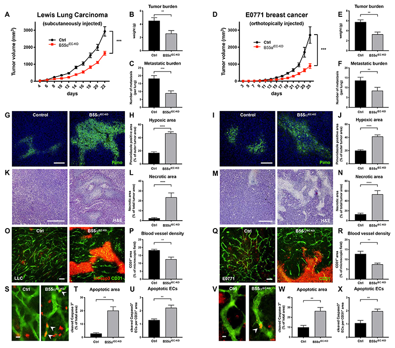
Figure 3. EC specific deletion of B55α leads to a strong delay in tumor growth lung metastasis, due to a reduced number of blood vessels.
(A-F) Tumor progression after EC specific deletion of Ppp2r2a following the subcutaneous injection of LLC cancer cells, showing delayed tumor growth, a smaller tumor burden and less lung metastasis in mutant mice (n ≥ 8 per group) (a-c) and orthotopically injected E0771 breast cancer cells (n ≥ 9 per group) (D-F).
(G-J) Representative images and quantifications of hypoxic areas in LLC (G,H) and E0771 tumors (n ≥ 8 per group, Scale bars = 100µm) (I,J).
(K-N) Representative images and quantifications and of necrotic areas in LLC (K,L) and E0771 tumors (n ≥ 7 per group, Scale bars = 400µm) (M,N)
(O-T) 70µm thick cryosections of LLC (O,S) and E0771 (Q,V) tumors stained for CD31+ blood vessels and the apoptosis marker cleaved Caspase3 and statistical analysis of blood vessel density (P,R), total area of apoptotic (tumor) areas (T,W) and cleaved Caspase3+ ECs (U,X) (n ≥ per group, Scale bars (O,Q) = 100µm; Scale bars (S,V) = 10µm).
P values are *p<0,05; **p<0,001; ***p<0,001; ****p<0,0001.
Graphs show standard error of the mean (SEM).

