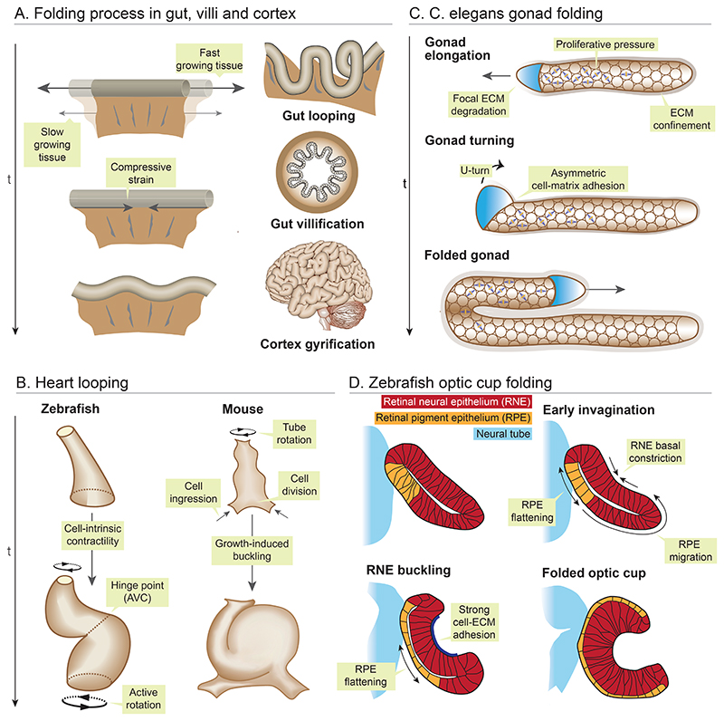
Figure 1. Physical principles of folding and looping morphogenesis.
(A) A faster growing tissue confined by slower growing adjacent tissue experiences compressive forces, which generates mechanical instabilities, driving brain cortex folding, mid gut lopping and gut villification. (B) In zebrafish, actomyosin forces twists the two chambers of the heart around the atrioventricular (AV) canal, resulting in torsion of the linear heart tube into S-shape. In mice, asymmetric rotation, preferential cell ingression and proliferation at the ventral pole buckles the linear heart tube rightwards to form a loop. (C) In C. elegans proliferative pressure from germ cells and ECM confinement propels gonad elongation, while direction of folding is driven by polarised cell-ECM interaction. (D) Optic cup morphogenesis in zebrafish is a multifaceted process. RPE cells migrate and get incorporated into the inner RNE layer, which compresses and buckle the RNE. The basal surface of the RNE layer constricts and is constrained by ECM adhesion which further accentuates the buckling process. The outer RPE layer stretches and flattens to drive the optic cup folding.

