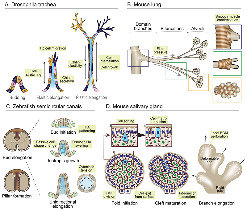
Figure 3. Morphogenesis of epithelial buds and branches.
(A) The drosophila trachea branches through epithelial remodelling in post-mitotic cells. Buds elongate through active migration of tip cells, which increase tension in the stalk, leading to elastic cell stretch and branch outgrowth. Area growth and intercalation of stalk cells enables plastic elongation, restricted by elasticity of a lumenal chitin matrix. (B) Domain branches, bifurcations and alveoli in the mouse lung are sculpted by smooth muscle fibres which locally condense at branch tips and form a physical growth barrier. The airway epithelium buds between smooth muscle fibres under positive hydraulic force. (C) Zebrafish semi-circular canals appear as buds extending into the otic vesicle lumen. Hyaluronic acid (HA) is secreted locally beneath the prospective bud, and osmotically swells, leading to budding through passive cellular deformations. Cytocinches between cells increase circumferential tension, translating isotropic HA swelling into anisotropic bud elongation. (D) The mouse salivary gland buds through folding of the epithelial surface and basement membrane. Cells exit the surface layer (blue nuclei) and divide in the core of the bud (green nuclei). Daughter cells (blue cell) then sort from inner cells owing to differential adhesion, return to the outer, and restore adhesion with the basement membrane. Growth of the surface layer folds the basement membrane, forming clefts stabilised by fibronectin secretion. Outgrowth of new branches is aided by focal ECM degradation, converting it from a stiff shell to a deformable lattice.

