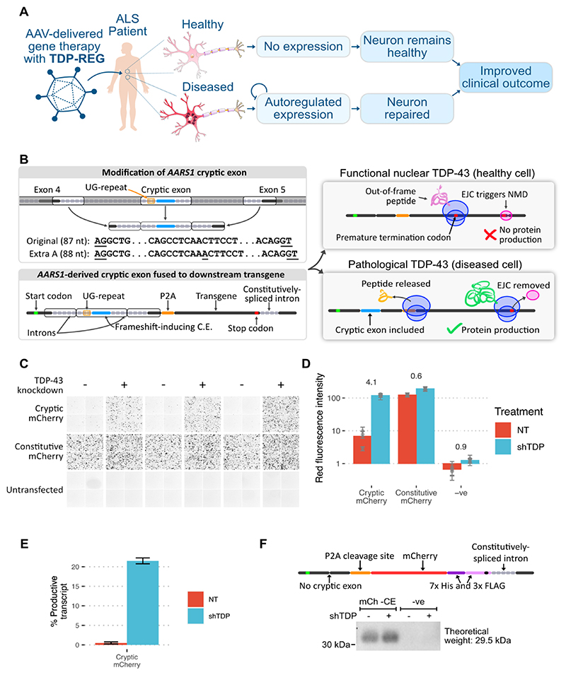
Fig. 1. Regulatory upstream CE controls downstream transgene expression (TDP-REGv1).
A: Schematic showing the intended function and purpose of this work. B: Schematic of TDP-REGv1. Top left: the genomic locus surrounding the human AARS1 CE region was modified, with the middle parts of the introns removed to reduce size, and an extra adenosine added to the CE sequence to enable frame-shifting. The AG and GT splice sites of the CE are underlined. Bottom left: the modified sequence from part A was incorporated into a “minigene”. Top right: when the CE is excluded, the start codon is out-of-frame with the transgene, triggering nonsense-mediated decay (NMD) due to the downstream exon junction complex (EJC). Bottom right: when the CE is included, the transgene is in-frame with the start codon, resulting in protein expression. C: Fluorescence microscopy images (red channel) showing SK-N-BE(2) cells with (shTDP) or without (NT = “not treated”) TDP-43 knockdown, transfected with a vector fusing the upstream AARS1-derived sequence to mCherry (“cryptic mCherry”) or a constitutive vector. D: Quantification of the images in Part C; numbers show log2-fold-change in TDP-43 knockdown cells; each dot shows the average of one well (three wells per condition), with error bars showing the standard deviation within each well (four per well). E: Summary of Nanopore sequencing results for the cells in Part C; error bars show standard deviation across three replicates. F: Top: schematic of mCh -CE vector, without the CE but with a downstream His/Tri-FLAG dual tag to enable sensitive detection. Bottom: Western blot of cells transduced with the above vector (“mCh -CE”) or a completely different vector (“-ve” - a Prime Editing vector). Samples were enriched with a His-tag pulldown, then blotted with an anti-FLAG antibody.

