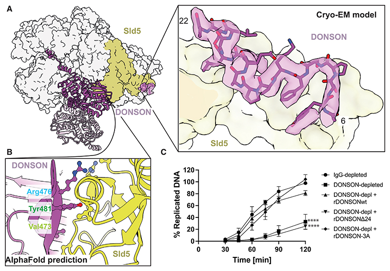
Figure 3. DONSON binding to Sld5.
(A) One isolated CMG from the dCMGDo structure, engaged by dimeric DONSON. Only one DONSON protomer binds one CMG. The zoomed in view shows the N-terminal tail of DONSON, tethered to the DONSON core via an unstructured (invisible) linker domain, and bound to Sld5.
(B) AlphaFold prediction of the engagement interface between the DONSON globular dimerization domain and the Sld5 B-domain.
(C) DNA replication reaction was set up in IgG-or DONSON-depleted extract optionally supplemented with 16 nM recombinant wild-type or indicated DONSON mutants. The synthesis of nascent DNA was followed by incorporation of α 32P-dATP into newly synthesized DNA at indicated times. n = 3, mean with SEM presented. Two-way ANOVA comparing with IgG-depleted, **** p < 0.0001.

