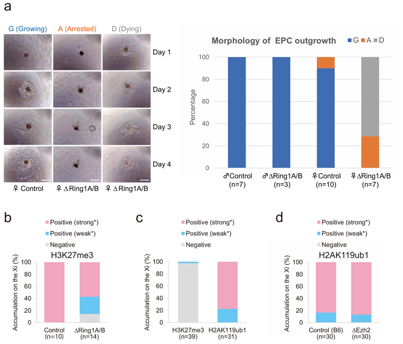
Extended Data Fig. 4. Morphology of ΔRing1A/B TGCs derived from E7.5 ectoplacental cone and quantification of H3K27me3 and H2AK119ub1 Xi accumulation.
a Female-specific lethality of TGCs upon Ring1A/B (PRC1) deletion. Morphology of EPC outgrowths, containing TGCs were recorded from days 1 to 4 of culture. Each TGC culture was categorized into three categories, Growing (G), Arrested (A), and Dying (D), based on their morphologies. Representative pictures are shown on the left. Scale bar: 100 μm. Summary chart is shown on the right. ΔRing1A/B female EPC outgrowth showed more severe phenotypes such as Arrested (A) and Dying (D) than male mutants and controls. b, Quantification of H3K27me3 Xi accumulation in control and ΔRing1A/B TGCs illustrated in Fig. 2d. c, Quantification of H3K27me3 and H2AK119ub1 Xi accumulation in nuclei from sections in Fig. 4b. d, Quantification of H2AK119ub1 Xi accumulation in ΔEzh2 TGCs as illustrated in Fig. 4d. b–d, All nuclei are analysed as positive (strong* or weak* expression as an Xi focus) or negative. Percentage and numbers of cells analyzed are given.

