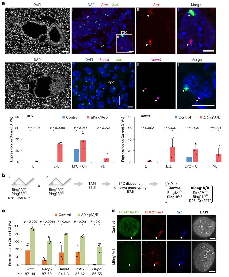
Fig. 2. Deletion of Ring1A/B (PRC1) results in loss of gene silencing on the Xi in extra-embryonic lineages.
a, X-linked gene expression on the Xi in extraembryonic tissues upon Ring1A/B deletion. Consecutive sections of ΔRing1A/B E7.5 embryo (Δ2) showing transcripts of two X-linked genes (Atrx and Huwe1) and Xist by RNA FISH. Top: Atrx and Xist. (i) Boxed area showing embryo proper (e), visceral endoderm (ve) and TGC. TGC has endoreplicated genome DNA and shows multi-spot RNA FISH signal (here, Atrx). (ii) Boxed region in i showing a TGC with biallelic Atrx expression (Xa and Xi). Middle: Huwe1 and Xist. (i) Boxed area showing ectoplacental cone (epc). (ii) Boxed region in i showing a cell with biallelic Huwe1 expression (Xa and Xi). Three independent embryos (Δ1: OR1–7, Δ2: OR1–8 and Δ3: OR8–15) were examined, and all showed similar results. Δ1 is shown in Extended Data Fig. 3b. Bottom: quantification of Atrx and Huwe1 biallelic expression (Xa and Xi) in three ΔRing1A/B E7.5 embryos totally deprived of H2AK119ub1 on the Xi (Δ1, Δ2 and Δ3). Mean (percentage of nuclei) ± standard deviation (s.d.) from three independent embryos. P values were calculated using two-sided t-test. For the number of analysed cells, see Extended Data Fig. 3a. Scale bars: 100 μm (whole embryos) and 10 μm (enlarged images). Arrowheads: Xist-coated Xi. Arrows: Xa. ExE: extra-embryonic ectoderm. Ch: chorion. b, Schematic of the experiment to generate TGCs. Embryos were recovered at E7.5 after intraperitoneal tamoxifen (TAM) injection to pregnant mother at E5.5. EPC was isolated and cultivated for 3 or 4 days to derive TGCs (Methods). c, Deletion of Ring1A/B induces escape of X-linked genes in TGCs. Percentage of cells showing biallelic expression (Xa and Xi) of five X-linked genes in control and ΔRing1A/B TGCs. Mean (percentage of nuclei) ± s.d.; n indicates number of TGCs from three experiments. The expected ratio and variance of biallelic expression in the control and knockout were calculated assuming a binomial distribution, and the differences between them were evaluated by two-sided binomial test. d, Immuno-RNA FISH showing lack of H2AK119ub1 but accumulation of H3K27me3 on the Xi in ΔRing1A/B TGC. Arrowheads: Xist-coated Xi. Scale bar, 10 μm. For quantification, see Extended Data Fig. 4b.

