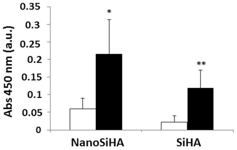
Figure 2.
Proliferation of EC2 endothelial cells on NanoSiHA and SiHA scaffolds without (white) and with (black) adsorbed VEGF after 7 days of culture. Cell proliferation was analyzed by measuring absorbance at 450 nm by CCK-8 protocol. *For each material, comparisons between conditions without and with VEGF. Statistical significance: *p < 0.05; **p < 0.01.

