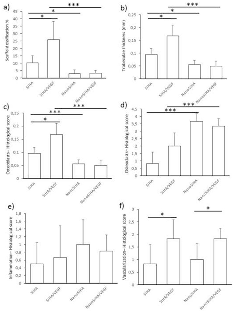
Figure 8.
Histomorphometrical studies for the different scaffolds implanted in osteoporotic sheep. (a) Ossification volume, (b) trabeculae thickness, (c) presence of osteoblasts, (d) presence of osteoclasts, (e) amount of inflammatory component and (f) vascularization degree. Statistical significance: *p < 0.05, **p < 0.01 and ***p < 0.005.

