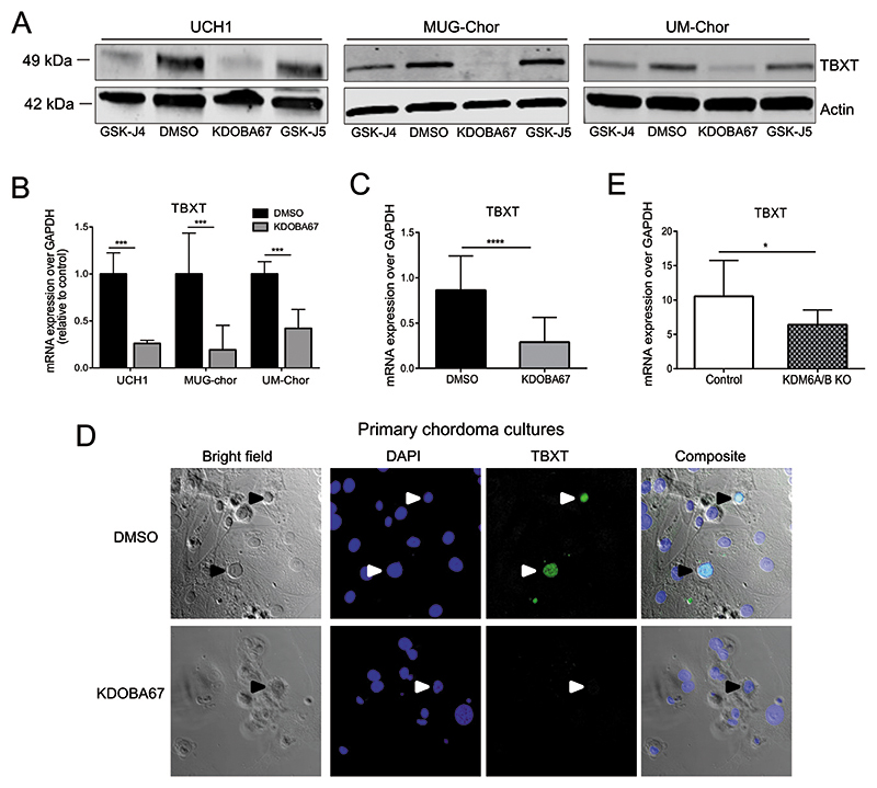
Figure 4. Inhibition of H3K27 lysine demethylase leads to inactivation of TBXT in chordoma.
(A) Protein expression of TBXT in chordoma cell lines, following treatment with KDOBA67 for 48 hours as assessed by western blotting. Beta actin is used as an endogenous control. Representative blots of 2 independent experiments. (B) TBXT expression in UCH1, MUG-Chor and UM-Chor following KDOBA67 treatment for 48 hours as assessed by qRT-PCR, in 3 independent experiments with 3 biological replicates per condition per cell line. (C) TBXT transcript level is reduced in primary chordoma cultures treated with KDOBA67 for six days, as assessed by qPCR. N=4 primary samples, at least three replicates per condition per sample. (D) Cell death and reduction in expression of TBXT (harrow heads) as shown by immunofluorescence in KDOBA67- and DMSO-treated patient-derived chordoma cultures for 6 days. TBXT-positive chordoma cells are interspersed between tumour-derived stromal cells. TBXT/green, DAPI/blue. 40X magnification. Representative pictures from one sample, experiment performed on 2 samples. (E) Expression of TBXT is reduced in UCH1 following double KO of KDM6A/B, assessed by qPCR. Quantification of two independent experiments, with two biological replicates per condition per experiment. *p ≤0.05, **p ≤0.01, ***p ≤0.001, ****p ≤0.0001.

