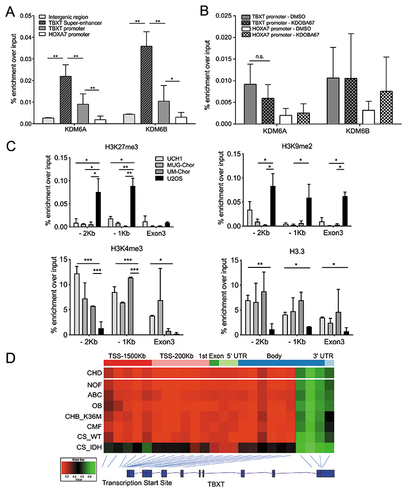
Figure 5. Histone modifiers epigenetically control TBXT expression in chordoma.
(A) ChIP-qPCR of KDM6A and KDM6B occupancy in UCH1 cells at the TBXT super-enhancer (indicated in Figure 3a), TBXT and HOXA7 promoter (-1Kb from TSS) and a control intergenic region (300Kb downstream of TBXT). Four biological replicates per condition. (B) ChIP-qPCR of KDM6A and KDM6B at the TBXT or HOXA7 promoter (-1Kb from TSS) in UCH1 cells treated with KDOBA67 or DMSO for 48 hours. Two biological replicates per condition per experiment, two independent experiments. (C) ChIP-PCR of H3K27me3 and H3K9me3 (top), H3K4me3 and H3.3 (bottom) in three chordoma cell lines (UCH1, MUG-Chor, UM-Chor) and the osteosarcoma cell line U2OS. Two independent experiments, with three biological replicates per condition per experiment. (D) The DNA at the promoter region of TBXT is hypomethylated in all primary human chordomas (n=35) and in other mesenchymal tumours not associated with the expression of TBXT. From top: chordoma, non-ossifying fibroma (n=12), aneurysmal bone cysts (n=9), osteoblastoma (n=12), chondroblastoma harbouring H3.3-K36M mutation (n=17), chondromyxoid fibroma (n=25), chondrosarcoma WT (n=2), chondrosarcoma-harbouring an IDH mutant (n=3). The average beta value for each probe is plotted with the position of the probe shown relative to the TBXT gene body. *p ≤0.05, **p ≤0.01, ***p ≤0.001, ****p ≤0.0001.

