Figure 3.
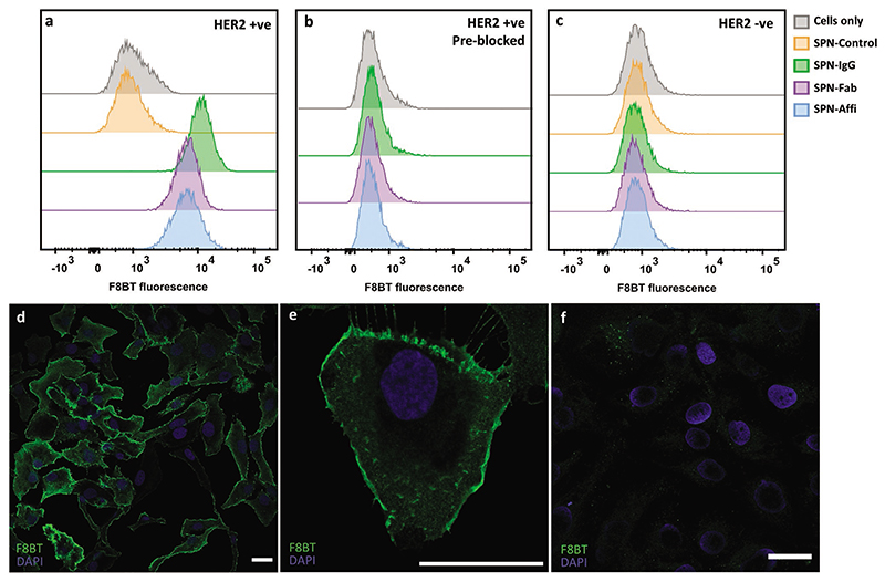
Targeted SPNs in vitro. Representative histograms from flow cytometry of a) SKOV3 cells (HER2 positive), b) SKOV3 cells (preblocked with free IgG/affibody) incubated with targeting and control SPNs, c) MDA-MB-468 cells (HER2 negative) incubated with SPNs. d,e) Confocal images of SKOV3 cells incubated with SPN–Affi. f) Confocal images of SKOV3 cells incubated with SPN-Control. Scale bars = 30 μm.

