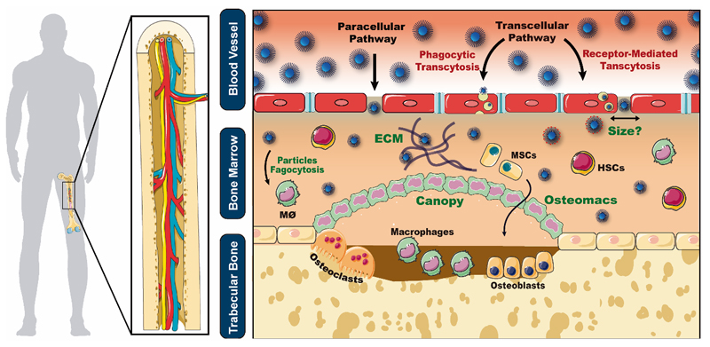
Figure 1.
Schematics of nanocarriers biological barriers and routes of extravasation from the sinusoidal vasculature into the extravascular space. Green text represents major barriers to nanocarrier delivery to bone cells (bone marrow ECM, macrophage-mediated phagocytosis, unknown fenestration size, bone canopy and its osteomacs - generally present in bone remodeling/regeneration stages).

