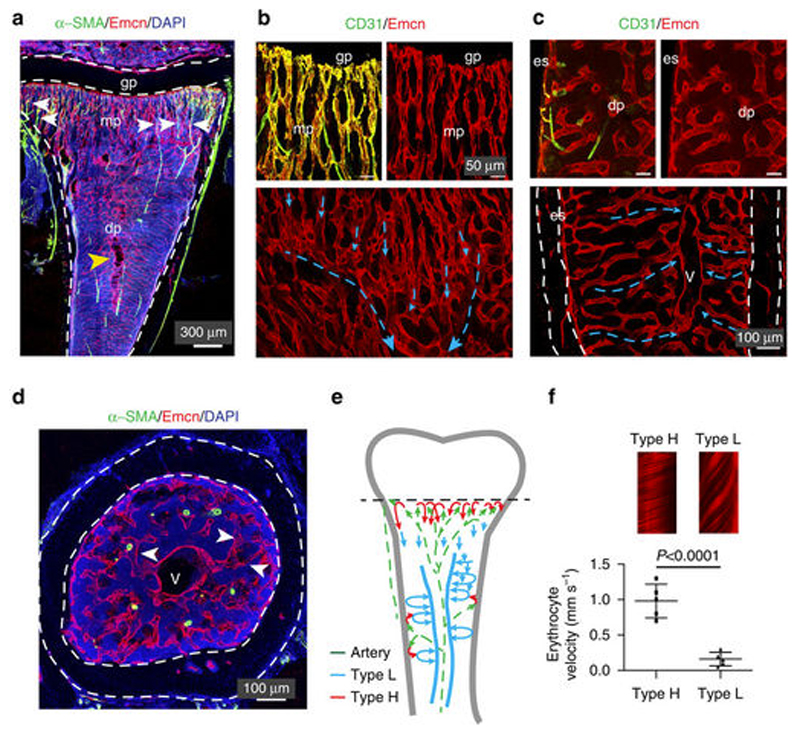
Figure 2.
Fluorescence microscopy imaging of 4-week-old tibial C57BL/6J mice vasculature. (A) Immunostaining of smooth muscle actin containing arteries (α-SMA positive cells, green channel), Endomucin (Emcn, red channel) and cell nuclei (DAPI, blue channel). mp- represents metaphyseal plate; gp- represents growth plate. White arrows indicate α-SMA cells connection to metaphyseal H-type vessels. (B, C) Confocal images proximal to the growth plate (b, top panel) or to diaphysis (c, top). CD31+ and Emcn-arteries terminate in type H vessels in metaphysis (CD31+ and Emcn+) and endosteum (es). No interaction with L-type vessels in diaphysis was observed. Blue arrows indicate blood flow from metaphyseal vessel columns (B) and endosteum (C), respectively. (D) Transversal tibial sections where sinusoidal L-type vessels (arrowheads) connect to the large central vein (v). Dashed lines represent compact bone. From these images, it is clear that CD31+ Emnc-arteries containing multiple smooth muscle cells cross the diaphysis. (E) Schematics of arterial (green arrows), H-type (red arrows) and sinusoidal/venous blood flow (blue arrows) of murin long bones. (F) Erythrocytes velocity data demonstrating the differences between type H and type L vessels. Adapted from [41] under Creative Commons Attribution 4.0 International License.

