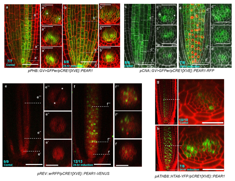
Extended Data Fig. 9. Overexpression of PEAR1 enhances the transcription of HD-ZIP III.
The transcription patterns of four HD-ZIP III, including PHB (a, b), CNA (c, d), REV (e, f) and ATHB8 (g, h) are visualized using their transcriptional fusion constructs. A longitudinal section is shown in the left panel, and the optical cross sections associated with this are shown in the right panels (the position of each section is indicated in the left panel). a-b, Transcription pattern of PHB (pPHB::GV>UAS::GFPer) in pCRE1[XVE]::PEAR1 plant before (a) and after 24 hours of induction of PEAR1 overexpression (b). PHB transcription is observed in whole vascular tissue at the initial and proliferative phase with peaks in xylem cells (a’), and its expression becomes concentrated into protoxylem cells (a” and a”’, asterisks indicate protoxylem cell). After the induction of PEAR1 overexpression, PHB expression in the central domain of the vascular tissue is maintained at the later stage, resulting in the radially symmetric PHB transcription pattern (b’ and b”). c-d, Transcription of CNA (pCNA::GV>UAS::GFPer) in pCRE1[XVE]::PEAR1-RFP plant before (c) and after 24 hours of induction (d). CNA transcription is observed mainly in xylem lineage at initial cells (c’), and becomes broader in whole vascular tissue, with peaks in procambial tissue, including PSE neighbouring cells (c”), and eventually its expression is gradually reduced in PSE and metaxylem, but is maintained in procambium, PSE neighbouring cells, as well as protoxylem cells (c”’). In a similar manner to PHB, CNA transcription in the central domain of the vascular tissue is maintained at the later stage when PEAR1-RFP is overexpressed (d”’). e-f, Transcription of REV (pREV::RFPer) in pCRE1[XVE]::PEAR1-VENUS plant before (e) and after 24 hours of induction (f). REV exhibits a distinct transcriptional pattern where its expression is initially uniform in vascular tissue (e’), and highest expression is localized in PSE, while decreasing towards xylem axis (e” and e”’). When PEAR1-VENUS is overexpressed (f), the transcription pattern of REV is also activated in the central domain of vascular tissue, resulting in the radial symmetric REV transcription pattern. g-h, The expression pattern of pATHB8::HTA6-YFP is highly specific to xylem cells (g), and its expression is enhanced after 24 hours of induction of PEAR1 overexpression with a broad expression domain (h). Number in each panel indicates samples with similar results of the total independent biological samples analysed.

