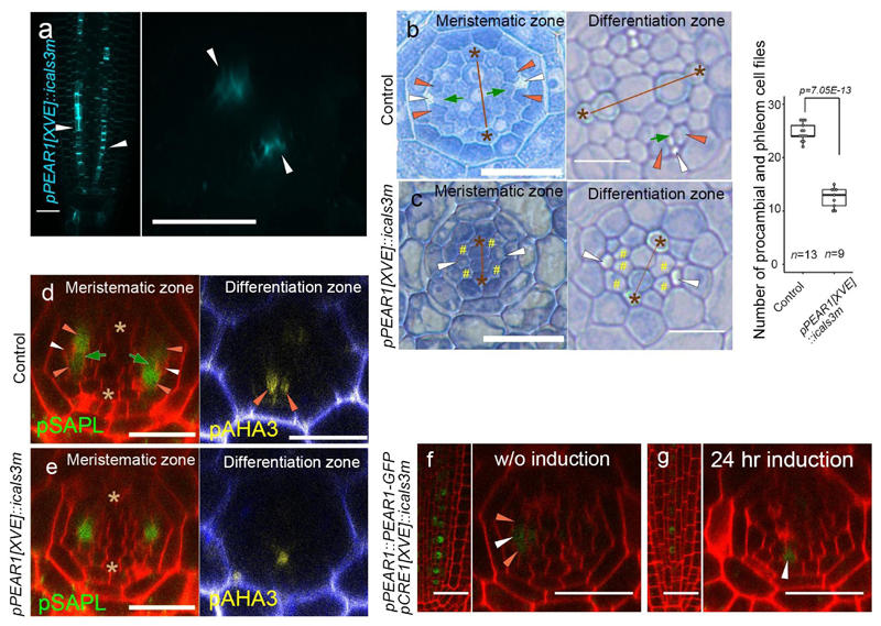
Extended Data Fig. 2. Inhibition of symplastic connection in early PSE results in the reduction of vascular cell number and in PSE-specific PEAR1-GFP localization.
a, Aniline blue stained primary root of pPEAR1[XVE]::icals3m after 24 hours of induction. Callose deposition occurs superficially in PSE cells (arrowheads, n=10). b-c, The vascular tissue of pPEAR1[XVE]::icals3m root, in non-induced (b, n=13) and after three days induction (c, n=9). In non-induced condition, PSE cells (white arrowheads) and their neighbouring cells, composed of MSE (dark-green arrows) and two lateral companion cells (orange arrowheads), are spatially separated from the xylem axis by intervening procambium. By contrast, after three days induction of callose deposition in PSE cells, only a single SE cell file is formed in each phloem pole (c, white arrowheads), and its neighbouring cells often touch the xylem axis (c, yellow hashtags). Number of procambial and phloem cell files is significantly reduced after three days induction. Boxplot centres show median. For more information on boxplots, see Methods. P value was calculated by two-sided Student’s t-test. d-e, Expression of Sister of APL (SAPL, AT3G12730) and ATPase 3 (AHA3, AT5G57350) in pPEAR1[XVE]::icals3m before (d, n=10 and 4, respectively) and after 24 hours of induction (e, n=10 and 4, respectively). SAPL is expressed in CC and MSE in meristematic zone, and AHA3 is expressed in differentiated CC (d). After induction, expression of these genes is restricted to a single cell file, indicating that symplastic cell communication between PSE and PSE-LN is required for the specification of PSE-neighbouring cell identity. f-g, PEAR1-GFP localization in pCRE1[XVE]::icals3m before (f, n=8) and after 24 hours of induction (g, n=7). PEAR1-GFP becomes specific to the PSE cell after the induction of callose deposition in whole vascular tissue, suggesting that PEAR1-GFP move in a short rage via plasmodesmata. White, orange and dark-green arrowheads indicate PSE, PSE-LN and PSE-IN, respectively. Asterisks indicate protoxylem (PX) cells. In a-g, n represents independent biological samples. Scale bars, 25 μm.

