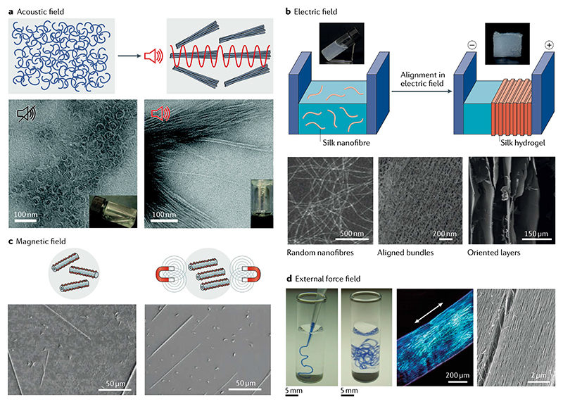
Fig. 3. External stimuli-induced oriented alignment of short peptide self-assembly.
a | Schematic (top) and scanning electron microscopy (SEM) images (bottom) of self-assembly and alignment of tripeptide (DFFD and DFFI) microfibrous structures by in situ ultrasonication48. b | Schematic (top) and transmission electron microscopy images (bottom) of the electric field-induced hierarchical alignment of silk nanofibres and the resulting anisotropic hydrogel49. c | Schematic (top) and SEM images (bottom) of magnetic field-induced alignment of the self-assembled diphenylalanine dipeptide nantotubes15. d | Noodle-like strings (left) obtained by injecting aqueous peptide amphiphilic (C15H31CO-VVVAAAEEE(COOH)) solution into phosphate-buffered saline. Uniform birefringence and aligned nanofibre bundles (right) induced by the shear force51. Part a is reproduced with permission from REF48, RSC. Part b is adapted with permission from REF49, Wiley-VCH. Part c is adapted from REF.15, Springer Nature Limited. Part d is adapted from REF.51, Springer Nature Limited.

