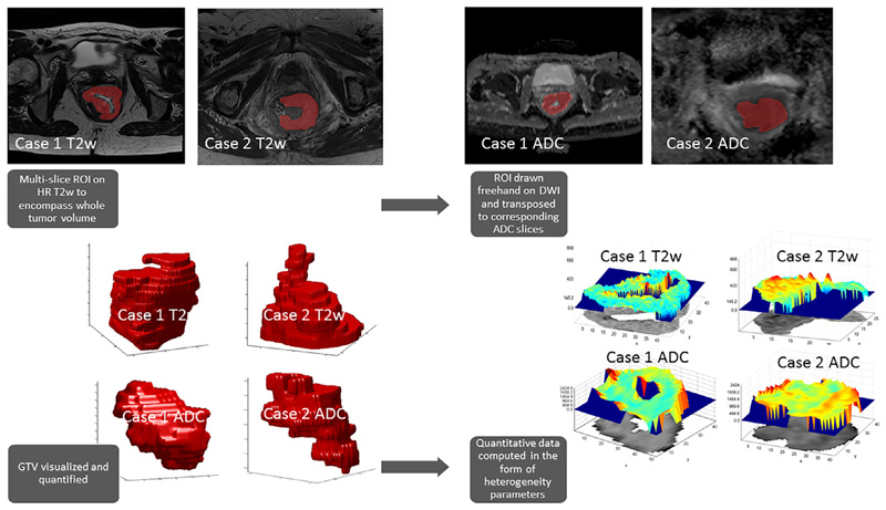
Fig 1.
Analysis workflow. Multi-slice regions of interest (ROI) were drawn around the tumor on high resolution T2w images as well as on Apparent Diffusion Coefficient (ADC) parametric maps with reference to high b value diffusion weighted images (DWI), generating a 3D whole tumor volume. From this volume, first-, second- and high-order statistical and fractal parameters were derived using in-house software. Both illustrated cases are of T4 anal canal tumors (Case 1 – Partial response post CRT, delayed recurrence; Case 2 – Partial response post CRT, subsequently CR and no recurrence at last follow-up).

