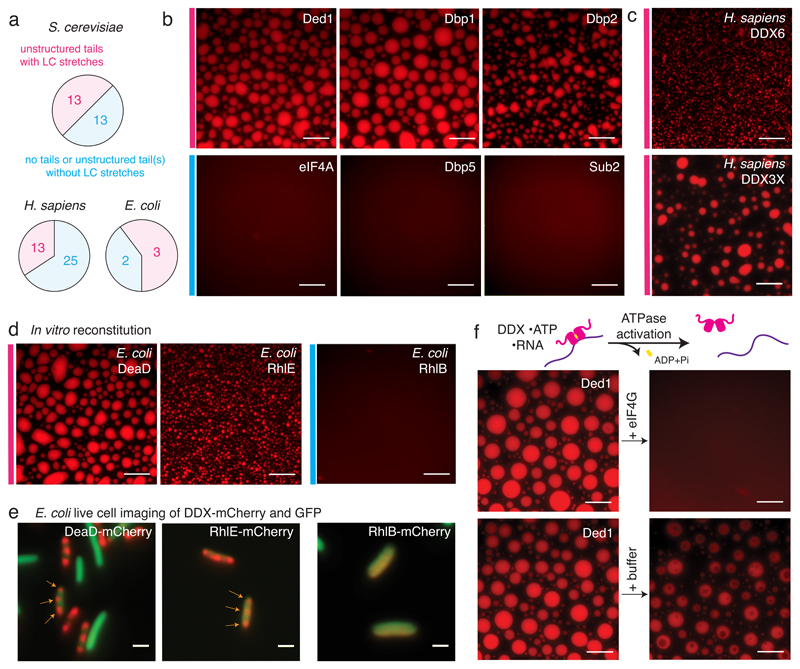
Fig. 2. Phase separation by DDXs is wide-spread and evolutionary conserved.
(A) Graph illustrating occurrence of LC domains in yeast, human and E. coli DDXs. (B-D) Representative images of at least 3 independent experiments: in vitro phase separation in the presence of ATP and RNA of select S. cerevisiae (B), human (C) and E. coli DDXs (D); scale bars 25 μm; for details see SI 2 Table 4. (E) Images of E. coli cells co-expressing mCherry-tagged DDXs and GFP. Subcellular DDX foci are marked with arrows. Scale bars 2 μm. (F) Droplets formed from Ded1-mCherry, ATP and polyU dissolve upon addition of recombinant eIF4GC-terminus, but not buffer. Scale bars 25 μm, representative images of > 3 independent experiments.

