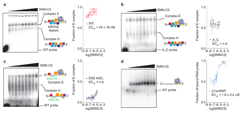
Fig. 4. Positive cooperativity between SMN-C5 and the splicing regulatory network.
a, Autoradiograph of the native gel showing the effect of SMN-C5 (from 10 nM to 2 μM) on the formation of early spliceosomal complexes in nuclear extracts. The free RNA probe, the H and E complexes are indicated. The colored circles on the scheme represent the splicing factors. The fraction of E complex was plotted as a function of the logarithm of the SMN-C5 concentration. b, Same experiment performed with the A-1C SMN2 E7 pre-mRNA fragment. The fraction of E complex was plotted as a function of the logarithm of the SMN-C5 concentration. c, Same experiment than a performed in the presence of the anti-sense oligonucleotide complementary to ESE2 (ESE2 ASO). The fraction of E complex was plotted as a function of the logarithm of the SMN-C5 concentration. d, Autoradiograph of the native gel showing the effect of SMN-C5 (from 800 nM to 250 μM) on the binding of in vitro reconstituted U1 snRNP on the SMN2 E7 pre-mRNA fragment. The positions of the free and bound RNA probes are indicated. The fraction of bound RNA probe was plotted as a function of the logarithm of the SMN-C5 concentration. For the four plots, experimental points and means are shown as plain and open symbols. The error bars represent the standard deviations. The experiment was performed four times (N=4) using the same batch of nuclear extracts.

