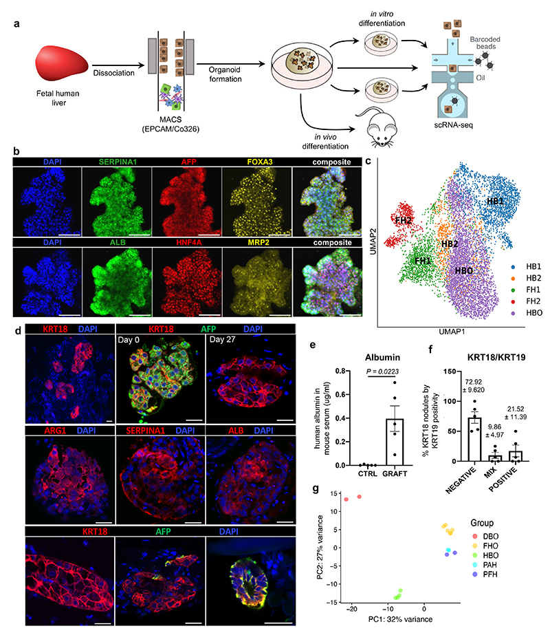
Fig. 4. Modelling early hepatic development in vitro using hepatoblast organoids.
a, Schematic representation of hepatoblast organoid (HBO) derivation and subsequent analyses. b, Immunostaining of hepatoblast markers in HBO grown in vitro; scale bars = 100 um. c, UMAP visualisation of fetal hepatoblast/hepatocyte differentiation stages along with HBO, confirming that HBO share the transcriptional profile of the HB2 stage of hepatocyte development. d, Immunostaining showing decrease of the fetal hepatocyte marker (AFP) in HBO after 27 days of engraftment while hepatocyte markers (KRT18, ALB, ARG1, and SERPINA1) were maintained, indicative of differentiation into mature hepatocytes. Immunostaining for biliary markers identified KRT19-positive cells in a subset of nodules, which organised into bile duct-like structures. Unless otherwise stated, pictures show grafts 27 days post-transplantation; scale bars = 20 um. e, ELISA analyses showing secretion human ALB in the serum of HBO recipient mice 27 days after engraftment (n=5 independent animals). f, Quantification (percentage) of KRT19-positive cells within KRT18-positive nodules. g, Principal component analysis (PCA) showing the divergence in gene expression profile between hepatoblast organoids (HBO; n=4 lines derived from 4 independent fetal livers), differentiated biliary organoids (DBO; n=2), fetal hepatocyte organoids (FHO; n=6), primary adult hepatocytes (PAH; n=2), and primary fetal liver (PFH; n=2). Data are presented as mean values +/- SEM; unpaired two-tailed t-tests.

