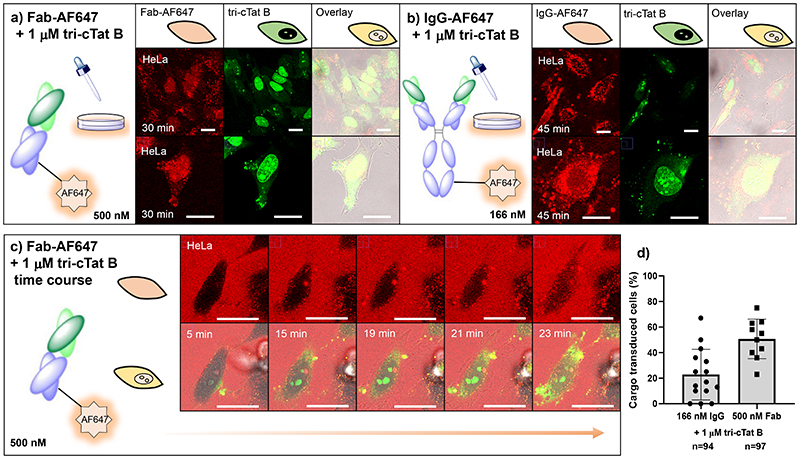
Fig. 4. Co-delivery of antibodies and antibody fragments in live HeLa cells using tri-cTat B.
(a, b) Post-wash live cell confocal microscopy images of HeLa cells treated with 500 nM mouse Fab fragment AF647 conjugate (Fab-AF647) and 1 μM tri-cTat B for 30 min (a) or 166 nM mouse IgG AF647 conjugate (IgG-AF647) and 1 μM tri-cTat B for 45 min (b) show homogenous distribution of Fab in cytosol and nucleus of cells with green nucleoli staining typical of tri-cTat B delivery; (c) Continuous live-cell confocal microscopy of trimers in HeLa cells treated with 500 nM Fab-AF647 (red) and 1 μM tri-cTat B (green); (d) quantification of the percentage of cells transduced with cargo (IgG-AF647 or Fab-AF647 - scored as positive when showing homogenous cytoplasmic and nucleolar fluorescence). Data presented as mean ± standard deviation. Scale bar: 20 μm.

