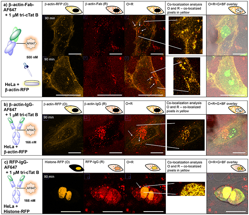
Fig. 5. Co-delivery of functional antibodies and antibody fragments in live HeLa cells.
HeLa cells were transfected with actin-RFP (a, b) or histone-RFP (c). 90 min post-wash live cell confocal microscopy images of cells treated with (a) 500 nM anti-β-actin mouse Fab-AF647 conjugate (β-actin-Fab-AF647) and 1 μM tri-cTat B for 30 min show co-localization of Fab (red) with actin stress fibres (orange); (b) 166 nM anti-β-actin mouse IgG2b AF647 conjugate (β-actin-IgG-AF647) and 1 μM tri-cTat B for 30 min show co-localization of antibody with actin stress fibres; (c) 166 nM anti-RFP mouse IgG1 AF647 conjugate (RFP-IgG-AF647) and 1 μM tri-cTat B for 30 min show co-localization of antibody with RFP fused to histone in the nucleus. G = green channel (tri-cTat B); O = orange channel (RFP fusion protein); R = red channel (AF647); Co-localization panel: co-localized pixels are shown as a mask of yellow pixels of constant intensity and all results shown present a significant correlation and are co-localized; BF = brightfield; scale bar: 20 μm.

