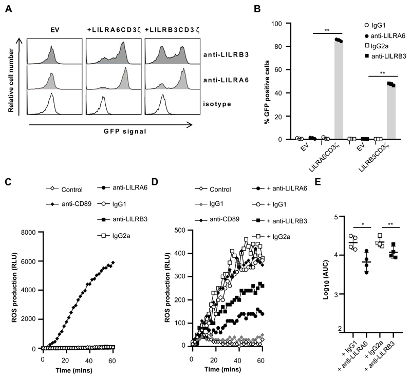
Figure 4. Cross-linking of LILRB3 suppresses FcαR mediated activation.
(A and B) GFP-expression in 2B4T cells expressing LILRCD3ζ (A6, B3) fusion proteins or empty vector (EV) control 2B4T cells following incubation at 37°C. Plates were previously coated with anti-LILRA6, anti-LILRB3 or isotype mAb. These mAb served as the agonist in these assays. Cells were incubated in coated wells for 18 hours, and GFP expression was measured by flow cytometry analysis. A representative experiment (A) and the integrated results from three separate experiments (B) were compared by Student t-test, where * = p < 0.05, ** = p < 0.01. (C) Cross-linking of LILRB3 does not induce ROS production by neutrophils. Neutrophils were incubated in the presence of 5 μg/ml FLIPr-like for 20 minutes, and then incubated on plates containing luminol. Plates were previously coated with anti-CD89, anti-LILRA6, anti-LILRB3 or isotype mAb. These mAb served as the agonist in these assays, and relative luminescence units (RLU) was measured over 60 minutes as an indicator of ROS production. (D and E) Cross-linking of LILRB3 suppresses CD89-mediated ROS production by neutrophils. Neutrophils (n = 4) were incubated on plates previously coated with anti-LILRA6, anti-LILRB3, IgG1 or IgG2a mAb. After 1 hour, neutrophils were stimulated through FcaR/CD89 by incubation on plates containing luminol that were previously coated with anti-CD89 or IgG1 isotype control. RLU was measured over 60 minutes as an indicator of ROS production. A representative experiment (D) and the integrated results from four separate experiments (E) were compared by Student t-test, where * = p < 0.05, ** = p < 0.01.

