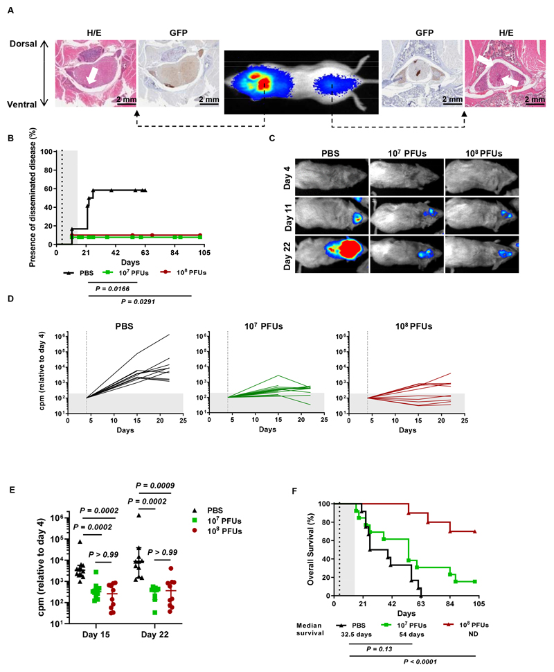
Fig. 4. Delta-24-RGD hinders the development of disseminated AT/RT lesions.
(A) Representative bioluminescence images of mice at 21 days after intraventricular administration of BT-12-luc/GFP cells showing the presence of luciferase signals at cranial and spinal locations. H/E and GFP staining demonstrated that the luciferase signals corresponded to local and disseminated tumor lesions. (B) Kaplan-Meier plot comparing the development of secondary AT/RT BT-12 tumors (log-rank test). Dots on the survival curves indicate death events. (C) Representative images of mice developing disseminated tumors acquired on days 4 (day of treatment), 11, and 22 after injection of BT-12-luc cells into the right ventricle. (D) Evolution of tumor growth measured as luminometry signals in mice treated with 107 or 108 PFUs of Delta-24-RGD or mock treated with PBS. The total counts per minute (cpm) measured in whole mice were normalized to the intensity of the signal on the day of treatment (dashed vertical line). (E) Analysis of differences in tumor sizes among the three experimental groups at days 15 and 22 after injection of tumor cells. Each dot represents an individual value, and bars indicate the median cpm ± 95% c.i. (Kruskal-Wallis test, Dunn’s correction). (F) Survival curve comparisons between mice bearing intraventricular BT-12 tumors that were mock treated (PBS) or treated with 107 or 108 PFUs of Delta-24-RGD (log-rank test). Dashed vertical lines in survival plots indicate administration of Delta-24-RGD.

