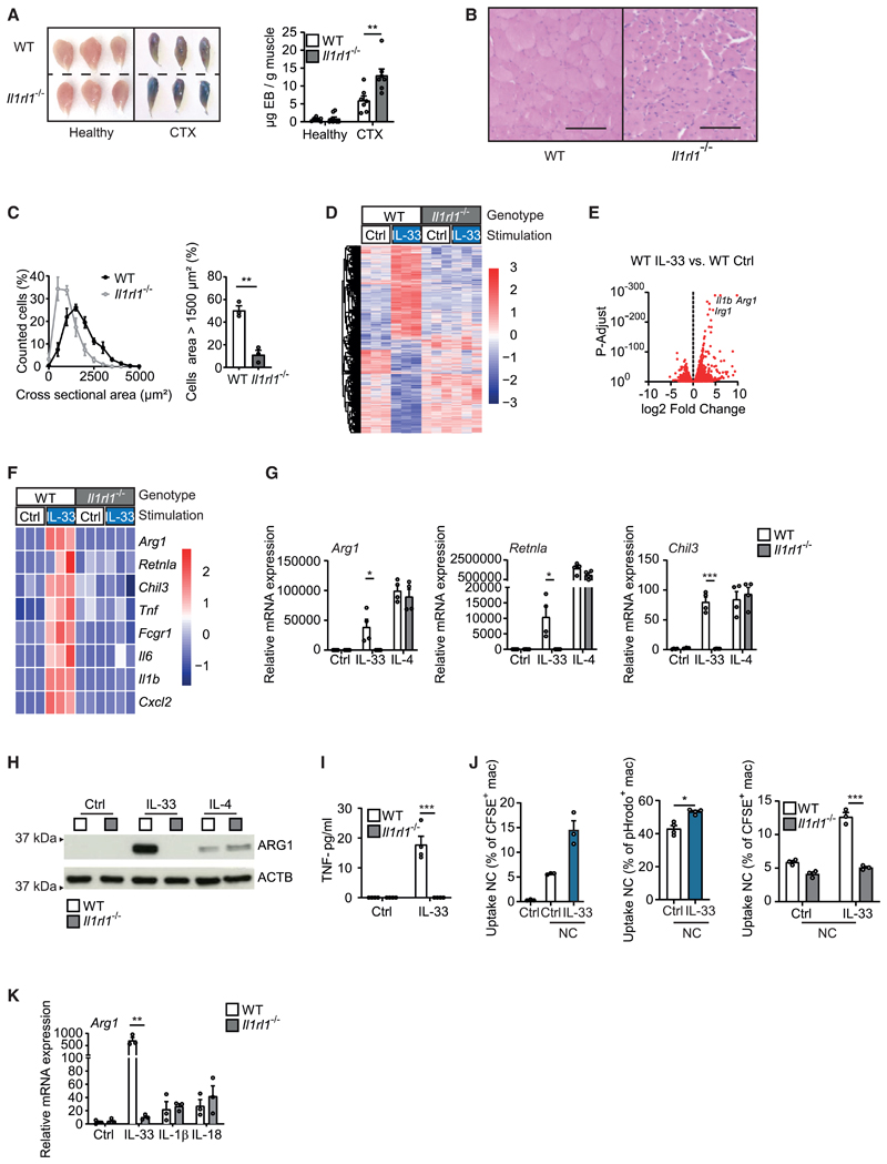
Figure 1. IL-33 promotes the resolution of injury-induced inflammation and the differentiation of AAMs.
(A) Determination of Evans blue accumulation in healthy and CTX-injected muscles of WT (n = 7) and Il1rl1−/− (n = 7) mice on day 7 after injury.
(B and C) Example H&E staining (B) and quantification and comparison of cross-sectional areas from muscle sections (C) of CTX-injected WT (n = 3) and Il1rl1−/− (n = 3) muscles on day 14 after injury. Scale bar indicates 100 μm.
(D–F) Bulk mRNA sequencing data of WT and Il1rl1−/− BMDMs that were treated with vehicle (Ctrl) or with IL-33 (10 ng/mL for 36 h). Data are presented as a heatmap illustrating differential gene expression (D), a volcano plot in which each dot represents a gene with a adjusted p < 0.05 (E), and a heatmap of genes encoding for alternative and classic macrophage activation markers (Z scores) (F).
(G–I) Quantitative real-time PCR (G), western blot (H), and ELISA (I) showing mRNA and protein expression of indicated markers of alternative activation (Arg1, Retnla, Chil3, ARG1) and classic activation (TNF) in BMDMs of WT and Il1rl1−/− BMDMs upon stimulation with vehicle (Ctrl) or IL-33 (10 ng/mL for 5 days) and IL-4 (20 ng/mL for 24 h). Data are representative of three individual experiments.
(J) Uptake of necrotic C2C12 cells (NC) after 1 h (CFSE-labeled) or 2 h (pHrodo-labeled) of phagocytosis. Data are representative of three individual experiments.
(K) Arg1 mRNA expression in response to IL-33 (10 ng/mL), IL-1β (10 ng/mL), or IL-18 (10 ng/mL) after 5 days of stimulation.
Data are presented as mean + SEM. *p < 0.05, **p < 0.01, and ***p < 0.001. See also Figures S1 and S2 and Video S1.

