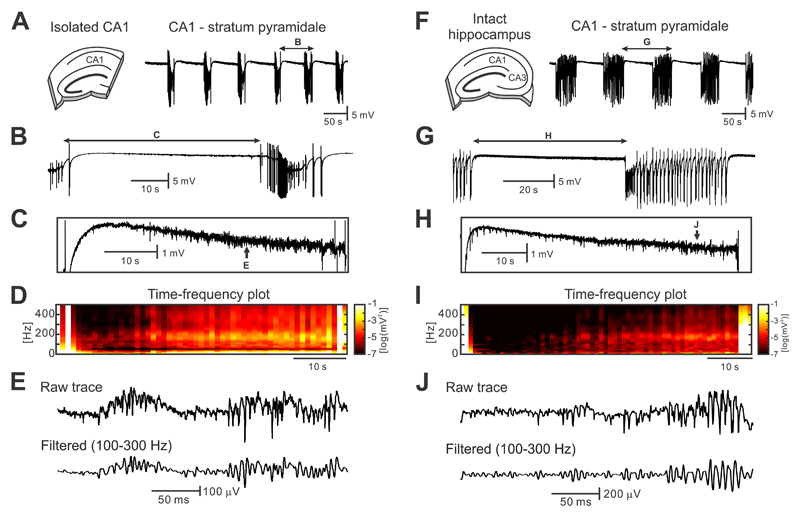
Figure 1. Seizures and interictal activity in the high-potassium model.
(a) In the isolated CA1, perfusion of ACSF with a potassium concentration of 8-10 mM leads to the development of spontaneous and repeated seizure-like episodes (n=114/17 seizures/slices). (b,c) Periods between seizures are characterized by the presence of HFA ~190 Hz with superimposed unit activity. These electrographic phenomena are accompanied by decreasing DC shift (n=27/9 interictal periods/slices). (d) Time-frequency map demonstrates the progressive increase in power in a high-frequency band with approaching seizure (n=114/17 seizures/slices). (e) Detail of interictal HFA. (f) In intact hippocampal slices, seizures are also generated within the CA1 region (n=83/15 seizures/slices). (g-i) Interictal periods in the intact hippocampus are characterized by the increase in amplitude and power of HFA (n=83/15 interictal periods/slices), the negative shift in DC potential (n=24/8 interictal periods/slices) and also by the presence of interictal discharges. (j) Example of interictal HFA in intact hippocampal preparation.

