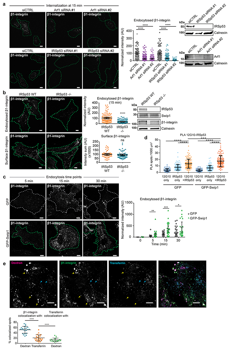
Extended Data fig. 5. Active β1-integrins are endocytosed via the CG pathway and CME.
(a) Representative micrographs and quantification of β1-integrin uptake in control- or IRSp53- or Arf1-silenced MDA-MB-231 cells. Representative immunoblots to validate Swip1 silencing. Scale bars, 10 μm. (b) Representative micrographs and quantification of endocytosed or surface (no endocytosis or acid wash) of murine β1-integrin (9EG7 antibody) in isogenic IRSp53−/− MEFs: IRSp53-KO-pBABE (-/-) and IRSp53-KO-pBABE-IRSp53 (WT) in which the expression of IRSp53 has been restored. Representative immunoblots of cell lysates blotted as indicated. Scale bars, 10 μm. (c) Representative micrographs of β1-integrin uptake in GFP- and GFP-Swip1-expressing cells and quantification of integrin uptake at the indicated times. Scale bars, 10 μm. (d) PLA with the indicated antibodies (from Figure 4i) in MDA-MB-231 cells expressing either GFP or GFP-Swip1. Plot shows the background controls for each. (e) Representative images and quantification of co-localization of β1-integrin-AF488 (12G10) with TMR-10 kDa dextran or AF647-transferrin after 1 min simultaneous uptake in MDA-MB-231 cells. Yellow arrows show regions of co-localization between β1-integrin and TMR-dextran and cyan arrows show regions where β1-integrin co-localizes with AF647-transferrin. Scale bars, 10 μm. For all plots, data are presented as mean values ± 95% CI. Statistical significance was assessed with two-sided Mann–Whitney tests, where n is the total number of cells pooled from 3 independent experiments (a-d) or from 2 independent experiments (e). (a) ****P<0.0001, (b) ****P<0.0001, ns = not significant, (c) *P = 0.0478, **P = 0.0058, (c) ***P = 0.0006, (d) ***P = 0.0001, ****P<0.0001, (e) ****P<0.0001. Number of analysed cells: (a) siCTRL, n = 103 cells, Arf1 siRNA #1, n = 177 cells, Arf1 siRNA #2, n = 157 cells, siCTRL, n = 100, IRSp53 siRNA #1, n = 142 cells, IRSp53 siRNA #2, n = 124 cells. (b) Endocytosed and surface β1-integrin, respectively: IRSp53 WT, n = 108 & n = 55 cells, IRSp53 KO, n = 100 & n = 60 cells. (c) GFP, n = 10, n = 22, n = 30 and n = 24 cells and GFP-Swip1, n = 15, n = 22, n = 32 and n = 26 cells, respectively. (d) For GFP, 12G10 only, n = 75 cells; IRSp53 only, n = 113 cells; 12G10 + IRSp53, n = 106 cells. For GFP-Swip1, 12G10 only, n = 78 cells; IRSp53 only, n = 92 cells; 12G10 + IRSp53, n = 111 cells. (e) β1-integrin-dextran, n = 22 cells; β1-integrin-transferrin, n = 22 cells and transferrin-dextran, n = 18 cells. Unprocessed blots and numerical source data are provided in Source data.

