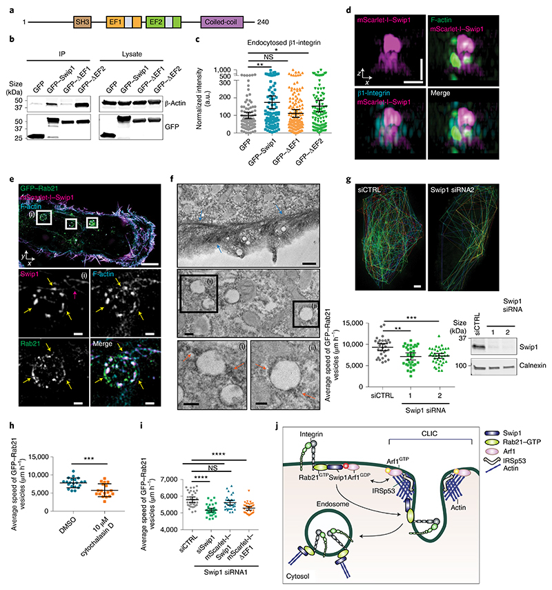
Fig. 6. Swip1–actin binding regulates integrin traffic.
a, Swip1 domains. EF, EF-hand domain containing a calcium-binding site (shown in grey); SH3, SRC homology 3 domain. b, Representative GFP-pulldowns of two independent experiments in HEK293 cells expressing GFP, GFP–Swip1 or truncated versions of Swip1 and blotted as indicated. IP, immunoprecipitation. c, Levels of β1-integrin uptake at 15 min in MDA-MB-231 cells expressing the indicated proteins. **P = 0.0034 and *P = 0.0356; n = 92 (GFP), 94 (GFP–Swip1), 161 (GFP–ΔEF1) and 101 (GFP–ΔEF2) cells. d, Representative SIM x–z projections of MDA-MB-231 cells expressing mScarlet-I–Swip1, immunostained for β1-integrin and labelled with phalloidin. Two independent experiments were performed. Scale bars, 0.7 μm. e, Representative SIM x–y image of MDA-MB-231 cells expressing mScarlet-I–Swip1 and GFP–Rab21 and labelled with phalloidin. The white squares highlight ROIs. The yellow arrows point to Swip1 overlap with actin on Rab21-containing vesicles. The pink arrow indicates actin filaments in close proximity to the vesicle. Scale bars, 5 μm (main image) and 0.5 μm (magnified views of (i); bottom). f, Electron microscopy images of GFP–Swip1 visualized using GBP-APEX. ROI (i) and (ii) are magnified (bottom). The arrows point to Swip1–APEX-positive patches adjacent to filament-like actin structures (blue) or vesicles (orange). Scale bars, 0.5 μm (top), 0.2 μm (middle and bottom left, magnified view of (i)) and 0.1 μm (bottom right, magnified view of (ii)). g, Average speed of Rab21 vesicles for each cell close to the TIRF plane over 2 min (bottom left). Representative tracks of Rab21 vesicles in a control or a Swip1-silenced cell (top) and representative immunoblot validating Swip1 silencing (bottom right). Scale bar, 5 μm; **P = 0.0005 and ***P = 0.0003; n = 31 (siCTRL), 33 (siSwip1 siRNA1) and 39 (siSwip1 siRNA2) cells. h, Average speed of Rab21-vesicle movement for each cell following treatment with cytochalasin D. ***P = 0.0007; n = 21 (DMSO) and 18 (cytochalasin D) cells. i, Average speed of Rab21-vesicle movement for each cell following Swip1 silencing and rescue with mScarlet-I–Swip1 or mScarlet-I–ΔEF1. ****P < 0.0001; n = 33 cells per condition. j, Swip1 directs integrins to CG-endocytosis. c,g–i, Data are the mean ± 95% CI; n is the total number of cells pooled from three independent experiments. Statistical significance was assessed using two-sided Mann–Whitney tests. Unprocessed blots and numerical source data are provided.

