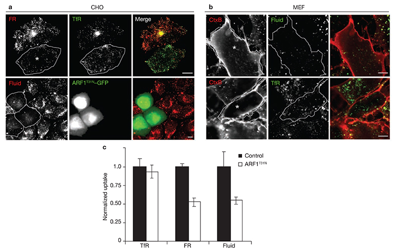
Figure 1. GDP-exchange deficient ARF1 inhibits uptake of GPI-APs and the fluid-phase.
(a) IA2.2 cells (CHO cells expressing FR-GPI (FR) and human TfR) were transiently transfected with ARF1T31N–GFP (outlined cells) for 18 h, and pulsed with Alexa568Mov19 Fabs and Alexa647 Tf (upper panels) or TMR–Dex (lower panels) for 10 min and processed for imaging. Images of internalized FR (red), TfR (green) and the fluid-phase (Fluid; red), are shown in grey-scale and colour merge. In transfected IA2.2 cells, the intracellular distribution of TfR containing perinuclear recycling compartment (REC) is altered, but Tf-uptake is unaffected. (b) MEFs transfected with ARF1T31N–GFP for 20 h, were pulsed with labelled probes for 5 min, fixed and imaged on a confocal microscope. Grey-scale and colour merge images of internalized Cy5–CTxB (CTx; red) with TMR–Dex (lower panel; green) or Alexa568Tf (upper panel; green) from a single confocal section are shown (transfected cells are outlined). In transfected cells, uptake of CTxB is blocked and fluid uptake is significantly reduced while TfR-uptake is unaffected. (c) Histogram showing uptake of TfR, FR-GPI (normalized to surface receptor expression level) and fluid-phase in ARF1T31N transfected cells plotted as a ratio to corresponding uptake measured in control cells. The error bars represent the weighted mean of fluorescence intensities ± s.e.m. (n = 61, 68, 100, 77, 63, 68; asterisks represent the cells transfected withARF1T31N). The scale bars in a and b represent 10 μm.

