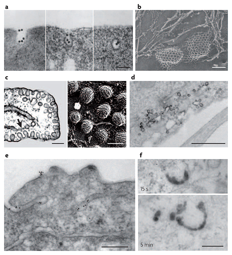
Figure 2. Electron micrographs of early intermediates in clathrin-dependent and independent pathways of endocytosis.
a|Thin-section views of reticulocytes incubated with gold-conjugated transferrin (AuTf) for 5 min at 37°C, which shows AuTf clustering into clathrin-coated pits and subsequently being endocytosed via coated vesicles. b | Rapid-freeze, deep-etch views of clathrin lattices on the inner surface of a normal chick fibroblast. c | Thin-section (left panel) and rapid-freeze, deep-etch images (right panel) of caveolae in endothelial cells. The arrow in the left panel points to the endoplasmic reticulum near deeply invaginated caveolae. d | Thin-section surface view of horseradish peroxidase (HRP)-conjugated cholera toxin in the process of internalization via grape-like caveolae in mouse embryonic fibroblasts. e | Thin-section images of green fluorescent protein (GFP) with a glycosyl phosphatidylinositol (GPI) anchor expressed in Chinese hamster ovary cells and incubated with gold-conjugated antibodies against GFP. The antibodies show putative sites for clathrin- and dynamin-independent endocytosis. These represent surface-connected tubular invaginations where the gold probe is concentrated with respect to the rest of the plasma membrane. f | Thin-section micrographs of internalized HRP-conjugated cholera toxin incubated at 37°C in mouse embryonic fibroblasts, showing early intermediates in clathrin- and caveolin-independent endocytosis. Clathrin- and dynamin-independent carriers (CLICs) are observed after 15 s (top panel) and GPI-anchored protein enriched early endosomal compartments (GEECs) are observed after 5 min (bottom panel). The scale bar in a and b is 100 nm, in c is 200 nm (left panel) and 100 nm (right panel), and in d–f is 200 nm. Part a reproduced with permission from REF. 101 © (1983) Rockefeller University Press. Part b reproduced with permission from REF. 102 © (1989) Rockefeller University Press. Part c reproduced with permission from REF. 103 © (1998) Annual Reviews. Parts d and f reproduced with permission from REF. 31 © (2005) Rockefeller University Press. Part e reproduced with permission from REF. 37 © (2002) Elsevier.

