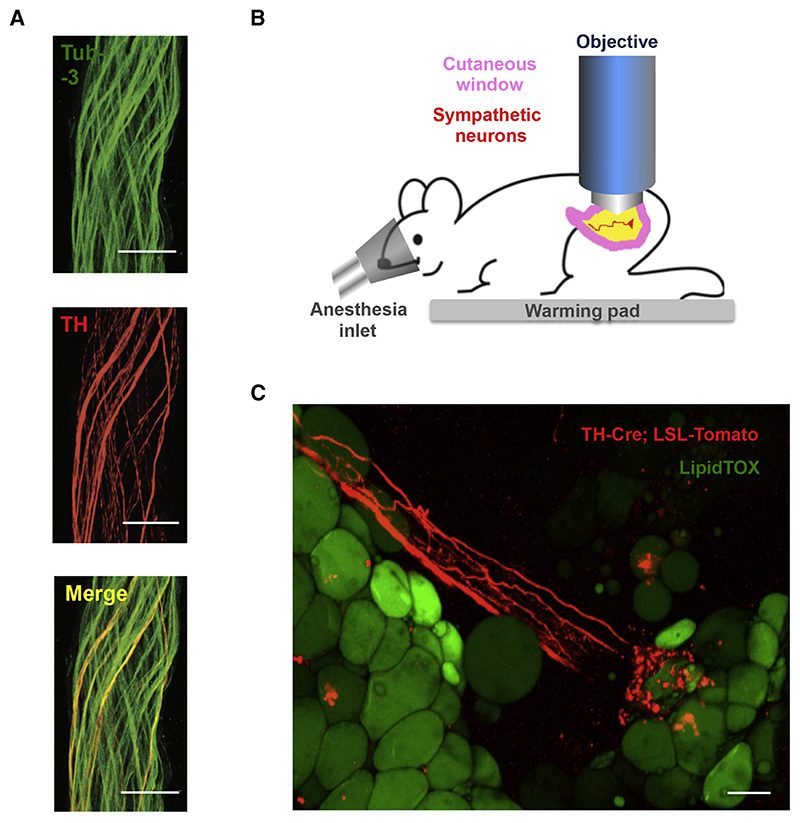
Figure 3. Catecholaminergic Neurons Innervating Adipocytes Integrate Nerve Bundles of Mixed Molecular Identity.
(A) Partial co-localization of TH (red), an SNS marker, and Tub-3 (green), a general PNS marker, shown by immunohistochemistry of nerve bundles dissected from the inguinal fat pads of WT mice. Scale bar = 50 μm. (B) Schematic representation of the two-photon intra-vital imaging of neurons in the inguinal fat pad. (C) Intra-vital two-photon microscopy visualization of a neuro-adipose connection in the inguinal fat pad of a live TH-Cre; LSLS-Tomato mouse – LipidTOX (green) labels adipocytes. Scale bar = 100 μm.

