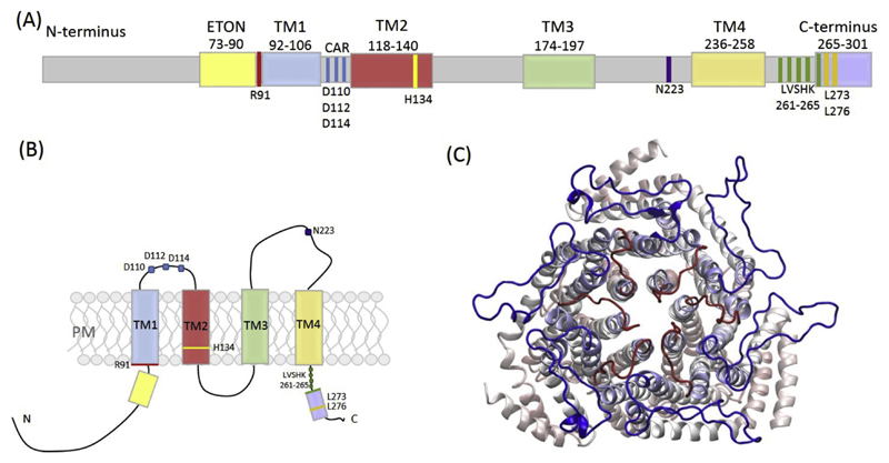
Fig. 1.
(A) Cartoon of full-length hOrai1 depicting the overall structure with important regions highlighted. (B) Schematic representation of a single hOrai1 monomer with the 4 transmembrane (TM) regions, N- and C-terminus and highlighted important residues like in (A). (C) Top view of a modelled hOrai1 hexameric structure showing TM1 (inner ring-pore), TM2-4, loop1 (red) and loop3 (blue) representing a closed channel.

