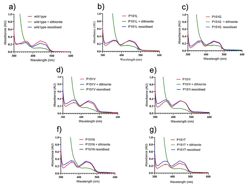
Figure 3. Spectral changes observed upon aerobic mixing of VAD with dithionite.
(a) Wild-type; (b) P151L; (c) P151G; (d) P151V; (e) (P151I; (f) P151N; (g) P151T. Protein and dithionite concentrations are 30 μM and 1.5 mM, respectively. The spectra measured before and immediately after the addition of dithionite, and after 10-minute incubation are in red, green, and blue, respectively. In all cases, the proteins are re-oxidized after 10 minutes.

