
Figure 8.
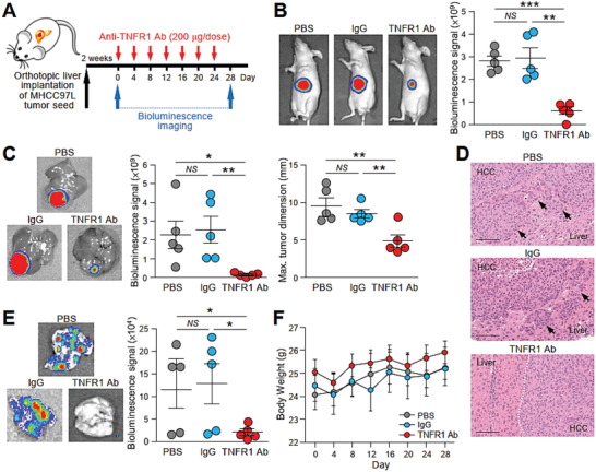
Treatment with TNFR1 neutralizing antibody effectively inhibits tumor growth and metastasis in mice implanted with metastatic tumor seed. A) Schematic diagram of the treatment regimen applied to mice implanted with luciferase‐labeled MHCC97L cells in the liver. Mice were administered PBS, IgG, or anti‐TNFR1 antibody (200 µg) via peritoneal injection every 4 days for 28 days (n = 5). B) Bioluminescence imaging of animals at the end of the experiment. Quantification of the luciferase signal is shown. The size of the liver tumors was measured and plotted. C) Ex vivo bioluminescence imaging of livers. Quantification of the luciferase signal is shown. D) Representative image of H&E staining of liver tissues showing the boundary of tumors obtained from (C). Dotted line indicates the bulging growth fronts of liver tumor. Arrows indicate the cluster of tumors nearby the liver‐tumor boundary. Magnification, 20 ×; Scale bar, 100 nm. E) Ex vivo bioluminescence imaging of lungs. Quantification of the luciferase signal is shown. F) Body weight of the mice was measured twice a week and plotted against time. Data are represented as the mean ± SEM; :p < 0.05, ::p < 0.01, :::p < 0.001 and NS, not significant from Student's t‐test.