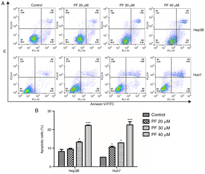
Figure 2.
PF induces HCC cell apoptosis. (A) Annexin V/propidium iodide (PI) staining of Hep3B and Huh7 cells after treatment with PF (20, 30 and 40 µM), as determined by fluorescence-activated cell sorting (FACS) analysis. (B) Quantitative assessment (%) of the apoptotic cells. The data represents the results from three independent experiment and expressed as mean ± SE. *P<0.05, ***P<0.001, significant variation relative to the control group. PF, Poncirus fructus; HCC, hepatocellular carcinoma.

