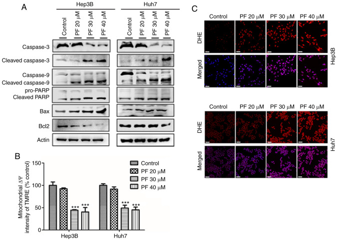
Figure 3.
PF induces HCC cell apoptosis by increasing ROS and disrupting mitochondrial membrane potential. (A) Western blot analysis was utilized to determine the levels of apoptosis-related proteins. (B) PF-induced changes in the mitochondrial membrane potential (ΔΨm) of Hep3B and Huh7 cells were determined with an ELISA reader after tetramethylrhodamine ethyl ester perchlorate (TMRE) staining. (C) The intracellular ROS levels in Hep3B and Huh7 cells after PF treatment. The cells were incubated with the ROS fluorescent probe, dihydroethidium (DHE), and evaluated using a confocal microscope. Scale bar, 30 µm. The data represents the results from three independent experiment and expressed as mean ± SE. ***P<0.001, significant difference with respect to the control group. PF, Poncirus fructus; HCC, hepatocellular carcinoma; ROS, reactive oxygen species.

