
Figure 2. ACE2-mediated inhibitory regulation of PVN neuronal perikarya.
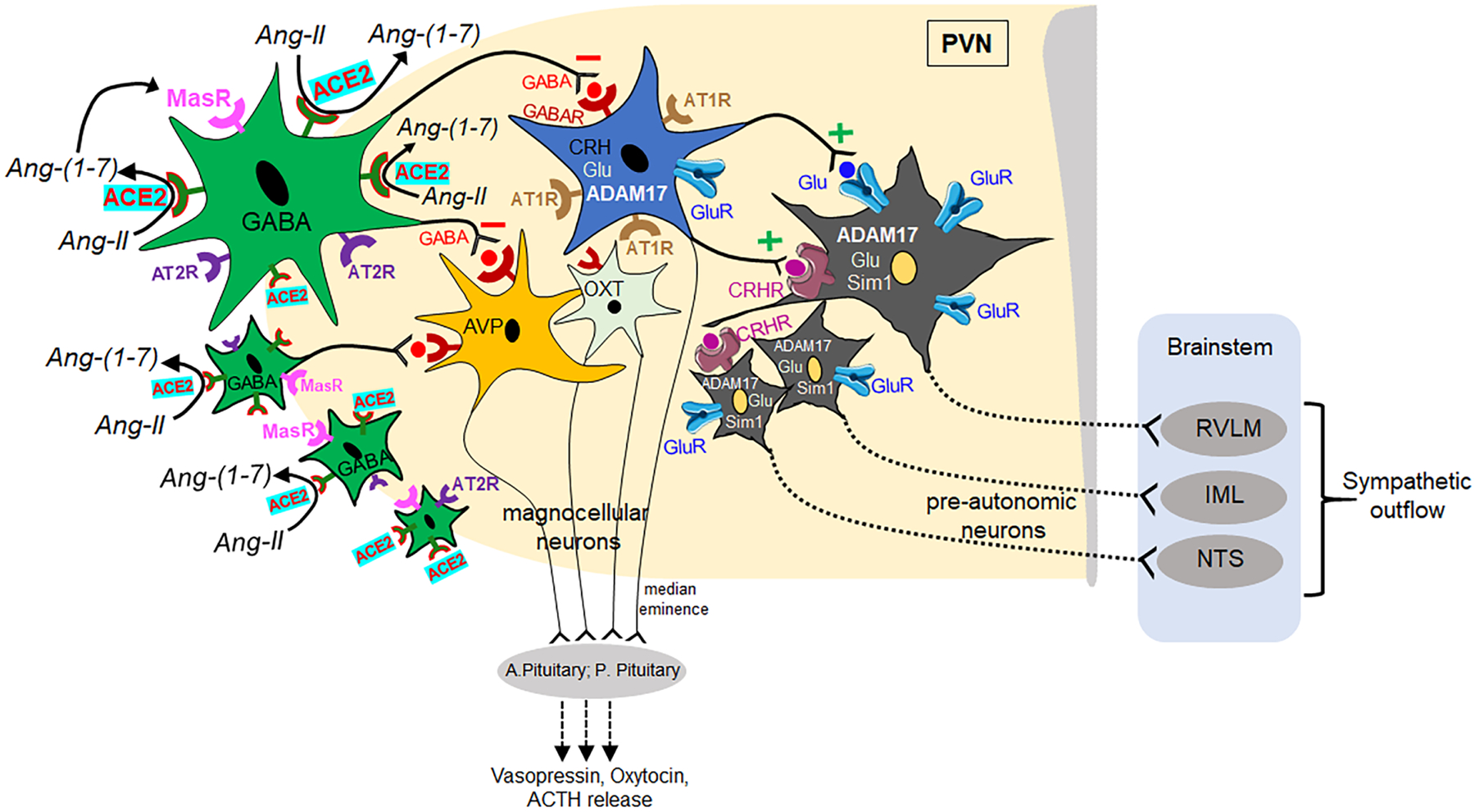
Diagram showing the ACE2-mediated inhibitory signaling in the PVN microcircuits. Stimulation of ACE2 activity in peri-PVN regions converts Ang-II into Ang-(1–7) and enhances GABAergic signaling to the PVN region that inhibits neuronal activity of autonomic and neuroendocrine functions. Upon stimulation, ACE2 blunts the activity of AT1R-containing neurons, which also contain CRH, ADAM17, Glutamate, and decreases the excitatory input to pre-autonomic neurons that project to the brainstem cardiovascular nuclei (RVLM, NTS) and IML, leading to decreased sympathetic outflow. Similarly, ACE2 also directly influences oxytocin and vasopressin neurons in the PVN and modulates their neuroendocrine release in the circulation. ACE2: angiotensin converting enzyme-2; AT1R: angiotensin II type 1 receptor; AT2R: angiotensin II type 2 receptor; CRH: corticotropic releasing hormone; Glu: glutamate; ADAM17: metallopeptidase domain 17; AVP: arginine vasopressin; OXT: oxytocin; CRHR: corticotropic releasing hormone receptor; ACTH: adrenocorticotropic hormone; sim1: single-minded 1; MasR: mas receptor; GABAR: GABA receptor; A. Pituitary: anterior pituitary; P. Pituitary: posterior pituitary; RVLM: rostral ventrolateral medulla; IML: intermediolateral column; NTS: nucleus tractus solitarius. Color coding: GABAergic neurons containing ACE2 (green), MasR (pink), AT2R (violet); Grey colored neurons represent pre-autonomic neurons containing ADAM17, glutamate and Sim1 along with GluR (blue), CRHR (violet).