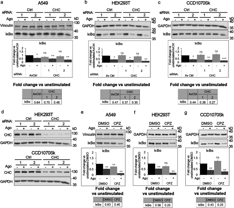
Fig. 6.
Blocking clathrin-dependent endocytosis enhances activation of canonical NF-κB signaling by LTβR. A549 (a), HEK293T (b, d) and CCD1070Sk (c, d) cells were transfected with siRNAs targeting clathrin (CHC) (two oligonucleotides) along with control, non-targeting siRNAs (two oligonucleotides) or treated with chlorpromazine (CPZ, e-g) along with DMSO, and stimulated or not with Ago for 1 h. Lysates of cells were analyzed by Western blotting with antibodies against the indicated proteins. Graphs show densitometric analysis of abundance of IκBα, normalized to loading controls (GAPDH or vinculin). Values are presented as a fold change vs unstimulated non-targeting controls – averaged non-targeting controls (AvCtrl) or DMSO, set as 1. Data represent the means ± SEM, n = 3 (a, b, f, g), n = 4 (c, e); ns - P > 0.05; *P ≤ 0.05; **P ≤ 0.01; ***P ≤ 0.001 by one sample t test. Tables present the fold change of IκBα abundance in stimulated vs unstimulated cells (means, n ≥ 3). d HEK293T and CCD1070Sk cells were analyzed with respect to the efficiency of clathrin knock-down. Representative blots are shown. The blots of GAPDH shown in panels b and c are also shown in panel d

