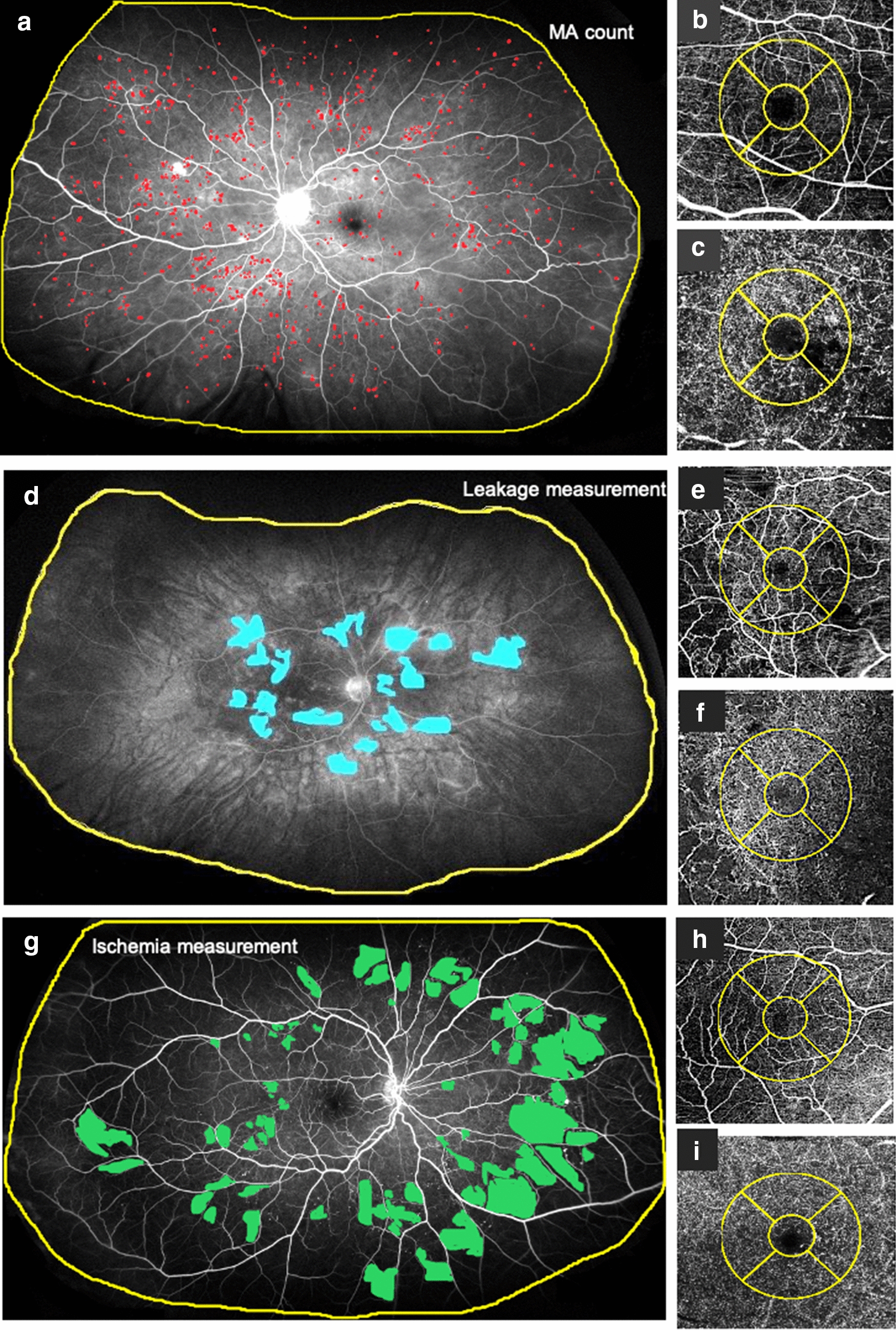
Fig. 2.

Ultrawide field angiography (UWFA) and optical coherence tomography angiography (OCTA) images of three diabetic patients. Patient 1 a–c: 60 years old female patient with severe DR. Patient 2 d–f: 49 years old female patient with moderate DR. Patient 3 g–i: 52 years old male patient with moderate DR. OCTA images shows the superficial (SCP) (b, e, h) and deep (DCP) (c, f, i) capillary plexus with the ETDRS Grid (yellow circle). The yellow line on the UWFA images (a, d, g) indicates the region of interest (ROI). a Microaneurysm (MA) analysis using an early phase UWFA image. Red dots indicate the MA-s. b OCTA image of the SCP of patient 1. c OCTA image of the DCP of patient 1. d Leakage analysis using a late phase UWFA image. Blue areas correspond to the leakage areas. e OCTA image of the SCP of patient 2. f OCTA image of the DCP of patient 2. g Ischemia analysis in an early phase UWFA image. The green field show ischemic areas. h OCTA image of the SCP of patient 3. i OCTA image of the DCP of patient 3
