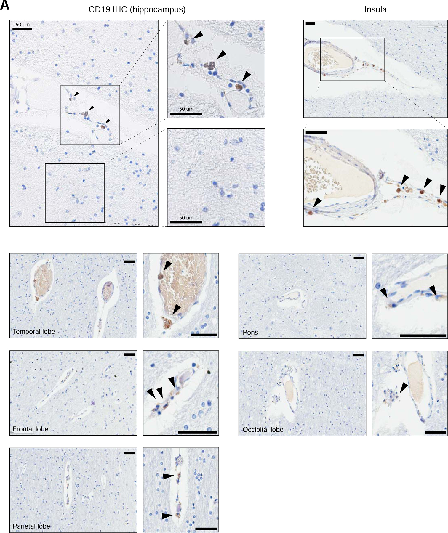
Figure 3. Perivascular staining of CD19 in human brain.

Representative immunohistochemistry staining for CD19 in human brain tissue. FFPE samples were stained for CD19 with a clinical protocol. Representative staining is shown for the hippocampus, insula, temporal lobe, frontal lobe, parietal lobe, pons, and occipital lobe. Scale bar = 50 µm. 5 slides were stained for the hippocampus, and 10 slides were stained for other brain regions.
