Figure 2.

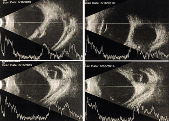
Ultrasound B-scan of both eyes showing abnormal optic nerve head with a retrobulbar hypoechoic lesion communicating with the optic nerve head

Ultrasound B-scan of both eyes showing abnormal optic nerve head with a retrobulbar hypoechoic lesion communicating with the optic nerve head