
Figure 1.
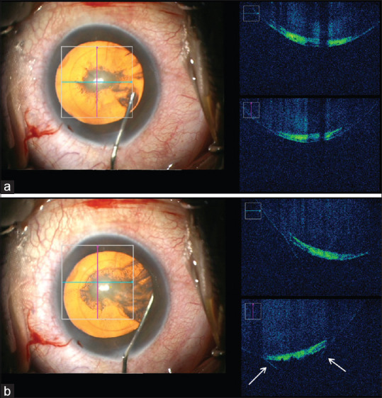
(a) Microscope-integrated intraoperative optical coherence tomography (MIOCT) focused on the posterior cortical-capsular interface. (b) Shows the initial stage of separation along with the posterior capsule and the cortical matter evident by posterior conical bulging of the posterior capsule (arrows)