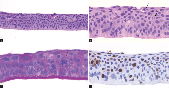
Figure 1.
Intraepithelial corneal neoplasia. (a) Histopathology demonstrates corneal full-thickness atypia, loss of cellular polarity, and dyskeratosis (eosinophilic cells). (Hematoxylin-eosin, original magnification 20×). (b) Higher magnification shows pleomorphic cells and frequent atypical mitoses (arrows) in the superficial layers of the epithelium. (Hematoxylin-eosin, original magnification 40×). (c) PAS stain highlights the abnormally thickened basement membrane and shows the atypical mitosis (arrow). (Periodic Acid Schiff, original magnification 40×). (d) Full-thickness epithelial proliferation is demonstrated by Ki67 nuclear cell proliferation marker (brown nuclei). (Mib-1 antibody, DAB chromogen, immunohistochemistry original magnification 40×)

