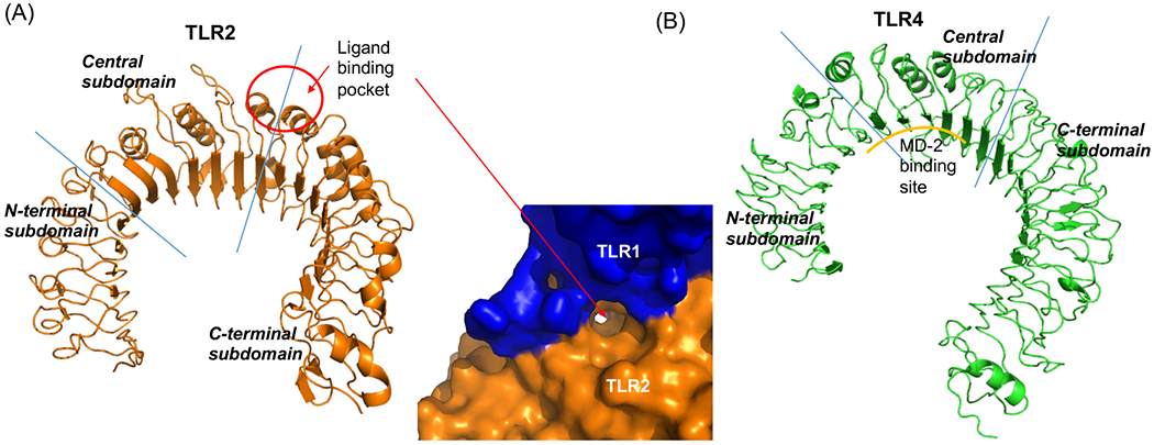
Figure 1. Horseshoe structure of TLR2 and TLR4.

Structures of (A) TLR2 (left, PBD: 2Z7X, TLR1/2 ligand Pam3CSK4 was removed from the original structure) and (B) TLR4 (right, PBD: 3FXI) were shown. Blue lines indicated the borders between N-terminal and central subdomains or between central and C-terminal subdomains. The red circle indicated the binding site of TLR2 ligands Pam3CSK4 and Pam2CSK4. In (A), The pocket in TLR2 dimerized with TLR1 was shown.
