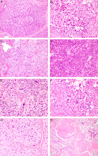
FIGURE 1.

Histologic features of MTC. A, Coagulative necrosis. B, Incipient necrosis defined as clusters of cells showing nuclear pyknosis, karryhorexis, and cytoplasmic condensation. C, Spindle cell morphology. D, Sheet-like growth. E, High nuclear grade. F, Multinucleation. G, Prominent nucleoli defined as nucleoli visible at ×100 magnification. H, Fibrosis/amyloid deposition >50% of tumor area (hematoxylin and eosin).
