
Figure 4.
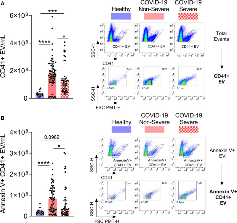
Platelet extracellular vesicles are released in patients with coronavirus disease 2019 (COVID-19). Circulating platelet extracellular vesicles (CD41+ extracellular vesicle [EV]) expressing phosphatidylserine or not were analyzed in plasma from healthy controls (n=18), patients with nonsevere (n=71) and severe COVID-19 (n=44). A, Total CD41+ EV were quantified (left) and representative scatter plots of CD41+ EV relative size and inner complexity are illustrated (right). B, Annexin V+ CD41+ EV were quantified (left) and representative scatter plots of AnV+CD41+ EV relative size are illustrated (right). The gating strategy is illustrated in Figure VIII in the Data Supplement. Samples with EV-concentrations close to the median of the whole population for each group were selected for representation. Data are represented as median with interquartile range (IQR). Statistical analysis: Data were not normally distributed (Shapiro-Wilk test). Kruskal-Wallis test with subsequent Dunn multiple comparisons test. FSC indicates forward scatter; and SSC, sideward scatter. *P<0.05, **P<0.01, ***P<0.001, and ****P<0.0001.