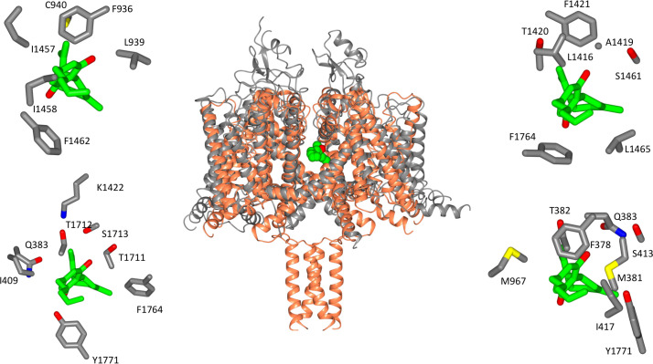
Figure 3. Location of CBD-binding sites in NavMs and the equivalent sites in hNav1.2.
(Centre) Structural alignment of the NavMs-CBD crystal structure (coral) and the hNav1.2 cryo-EM structure (grey). The RMSD of the aligned structures is 3.2 Å. (Top left): equivalent binding residues between domain I and domain II of hNav1.2 found within 4 Å of the CBD site. (Top right): equivalent binding residues in hNav1.2 between domains II and III located within 4 Å of the CBD-binding site. (Bottom left): Residues in hNav1.2 between domains III and IV within 4 Å of the CBD-binding site. (Bottom right): Residues in hNAv1.2 between domains IV and I within 4 Å of the CBD-binding site. In the surrounding panels the atoms in the protein are coloured by atom type, with carbons represented in grey, oxygen in red, nitrogen in blue, and sulphur in yellow, whilst the carbon atoms of the drug are depicted in green and the oxygens in red.

